Examples of Cryo-EM results
Lipid Nanoparticles
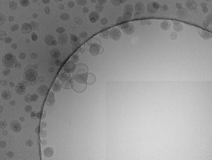
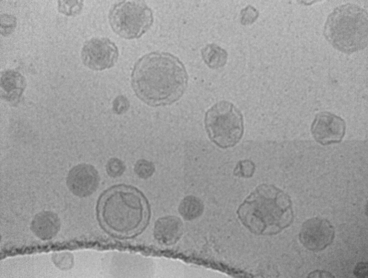
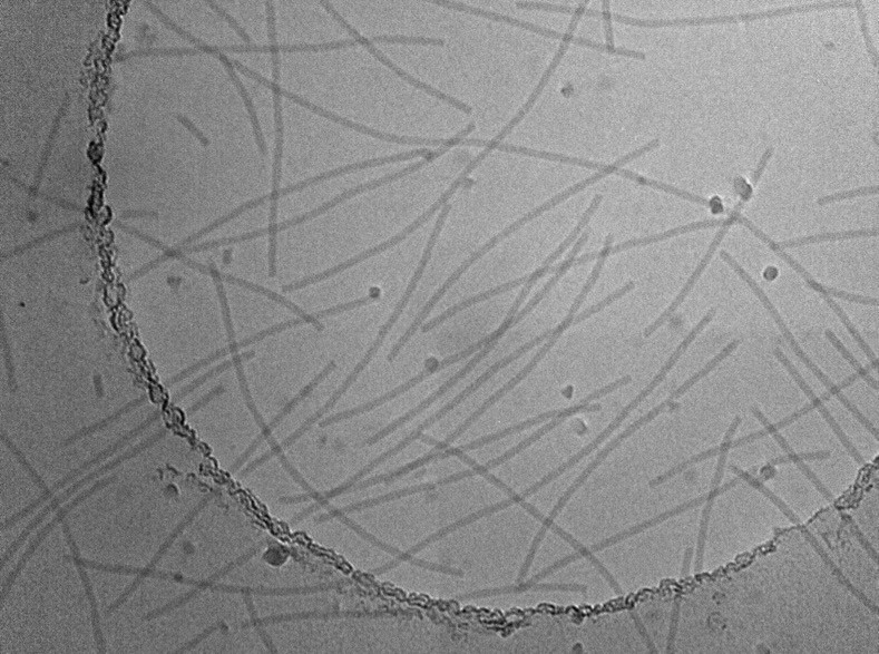
Size, morphology, lipid double layer, liposome contents... this is wath is studied in cryo-EM on the following lipid nanoparticle images.
Pseudoviral nanoparticles
Extracellular vesicles
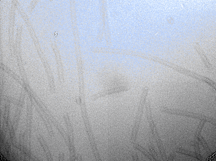
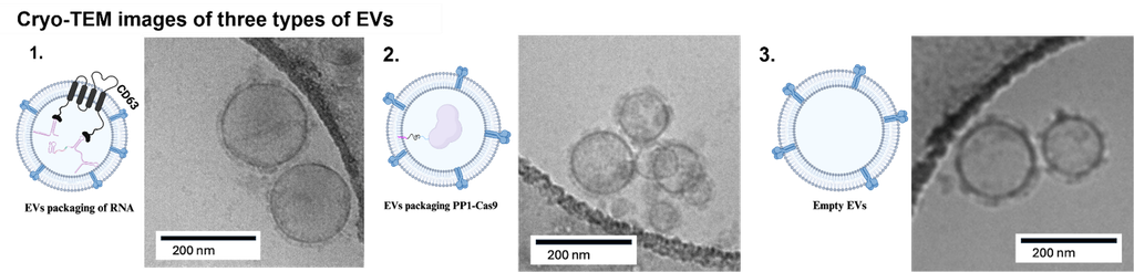
Cryo-TEM allows you to follow different steps in the development of extracellular vesicules (EVs) by visualizing their morphology and content, especially of the EVs that look empty, protein containing EVs and RNA-charged EVs. This work was realized in collaboration with Dr Tremblay's laboratory, the bioimagery platform and BIM Laboratory.
Gold Immunolabeling of VSV-G on Extracellular Vesicles
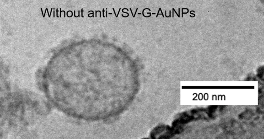
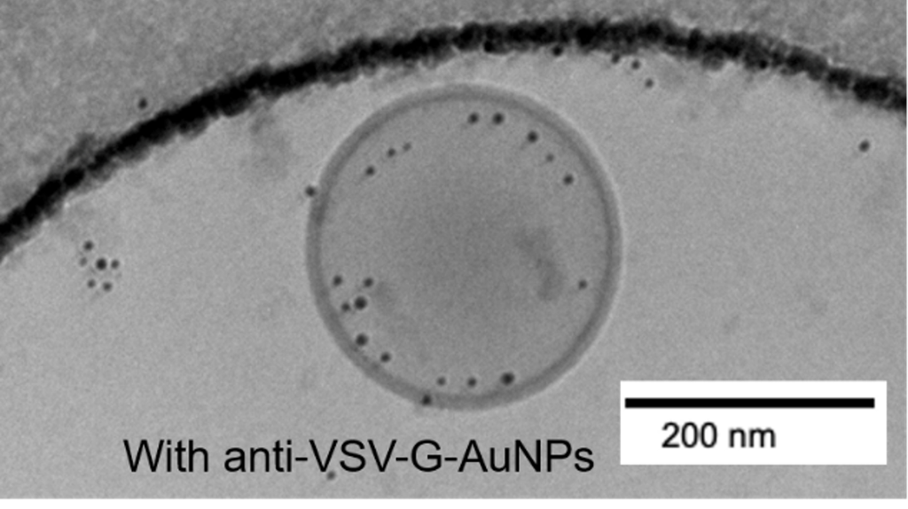
The incorporation of vesicular stomatitis virus glycoprotein (VSV-G) into extracellular vesicles (EVs) can enhance their stability, membrane fusion, and delivery of their contents. Cryo-TEM enabled the observation of the interaction between EVs, produced in collaboration with Dr. Tremblay’s laboratory, and 5- to 10-nm gold nanoparticles functionalized with PEG-Anti-VSV-G, developed at the BIM laboratory.
Research Project in Collaboriation with RQMEM
The Quebec Network of Electron Microscopy for Materials finances collaborative research projects between different nodes, includign projects between Université Laval node and McGill node.
Nanostructured and metalloproteined hydrogels : identification of observation conditions to measure in HRTEM and cryo-FIB
The complex interactions between biological hydrogels (e.g., mucus) and synthetic hydrogels (e.g., hydrogels for topical applications) and nanoparticles containing active ingredients (pharmacological, vaccine, or genetic) must be measured in several fields of health sciences (ophthalmology, immunology, cancer, etc.). However, visualizing the interactions between nanoparticles and hydrogels—whether biological or synthetic—poses a significant challenge in electron microscopy. First, hydrogels are fragile, water-saturated structures (more than 98%), and preparing them for observation under an electron beam requires specific techniques (e.g., cryosectioning followed by lyophilization for HRTEM observation; cryopreservation for cryo-FIB-SEM observation, etc.).
Since these organic samples are fragile, the instruments must be operated in modes, under conditions, and with settings (e.g., energy, focusing, magnification) that ensure low radiation doses. Furthermore, natural hydrogels such as mucus—found on the cornea, as well as the nasal, oral, gastric, intestinal, and vaginal walls, etc.—may contain high levels of metalloproteins. However, the concentrations of Fe, Mg, Mn, Cu, Ca, and Zn ions—the primary ions found in metalloproteins—must be sufficiently high to be detectable in sections of lyophilized or cryopreserved hydrogels examined by HRTEM or cryo-FIB-SEM. The concentration thresholds at which these metals would be detectable by EDS in such specimens must be measured.
In this project, synthetic hydrogels based on bovine mucins, which mimic the characteristics of certain human mucus, will be blended with lipid nanoparticles (representative of several drug delivery systems) and common metalloproteins (e.g., hemoglobin (Fe), hemocyanin (Cu), and zinc finger proteins). For HRTEM analysis, the hydrogels will be prepared via cryo-ultramicrotomy and lyophilized (U. Laval), while for FIB-SEM analysis, the hydrogels will be cryopreserved at McGill. The samples will be examined by HRTEM (including EDS) and by cryo-FIB-SEM. This project will enable the evaluation of all specimen preparation, dose, and signal constraints involved in the observation of this type of complex specimen.
This project's results will be communicated on RQMEM's website once the project is completed.