




A node is an institution of higher education that supports the Quebec Network of Electron Microscopy for Materials and where there is one or more microscopy platform. Some nodes are part of the institutions who originally submitted for funding with the Canadian Foundation for Innovation, in 2017, which are University of Sherbrooke, McGill University, Polytechnique Montréal, University of Montréal, Concordia University, Laval University and École de technologie supérieure. Other partner institutions can be added as RQMEM nodes with the approval of the RQMEM board of directors.
McGill
FEMR (Facility for Electron Microscopy Research) and MEMRG (McGill Electron Microscopy Research Group) are microscopy platforms members of the Quebec Network of electron microscopy for materials and bring together a diversified expertise with many microscopists collaborating with the Network.
FEMR is a platform that serves material science and biological science with expertise in tomography, molecular reconstruction and 3D vizualisation.
MEMRG holds the expertise for X ray quantification, materials and batteries analysis.
McGill Microscopists
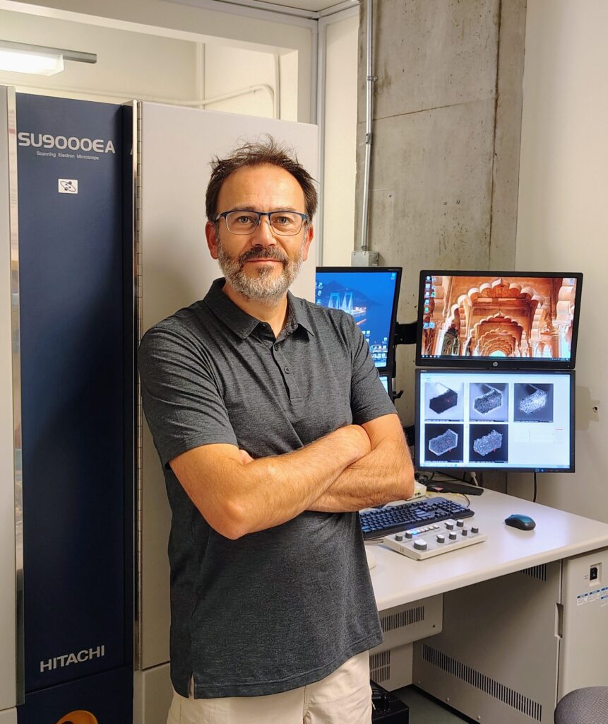
Nicolas Brodusch est actuellement Assistant de Recherche à l’Université McGill et est affilié au groupe de recherche sur la microscopie électronique de McGill (McGill Electron Microscopy Research Group, MEMRG, www.memrg.com), dirigé par le professeur Raynald Gauvin. En plus d’assurer la gestion opérationnelle des équipements et la supervision des projets de recherche des étudiants du groupe en lien avec le professeur, il a à cœur de développer des techniques innovantes, autant dans le domaine de la préparation d’échantillons que dans la caractérisation de matériaux divers par microscopie électronique à balayage.
Nicolas a obtenu un Diplôme Universitaire de Technologie (DUT), option Mesures-Physiques, à l’université Paris XI à Orsay, France, en 1997. Après avoir perfectionné ses techniques d’analyses et de recherches en tant que technicien de laboratoire dans divers laboratoires, il a obtenu un Master en Science et Génie des Matériaux par validation des acquis professionnels à l’université Paris-Saclay, Orsay, France, en 2018. Durant les seize années qu’il a passées au MEMRG, Nicolas a eu à cœur de pousser les limites des microscopes électroniques à balayage (MEB) sous sa supervision afin d’améliorer la résolution en imagerie et la qualité d’image mais aussi pour utiliser les détecteurs équipant ces instruments à leur plein potentiel. Ces travaux ont été reconnus internationalement par le biais de nombreuses publications scientifiques ainsi que par l’attribution d’un prix à la conférence Microscopy & Microanalysis à Hartford, CT, en 2014 pour l’article “Dark-field imaging of thin specimens with a forescatter electron detector at low accelerating voltage”, et plusieurs invitations en tant que présentateur invité à plusieurs conférences pendant les dernières années.
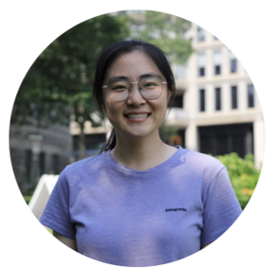
Wanting He is currently a postdoctoral fellow at McGill University and is affiliated with the Facility for Electron Microscopy Research (FEMR). RQMEM is proud to have access to her expertise in transmission electron microscopy (TEM), electron energy-loss spectroscopy (EELS), electron tomography, and nanomaterials characterization (i.e. plasmonics materials).
She received her B.S. in Materials Science and Engineering from Beihang University and her M.S. in Materials Engineering from the University of Southern California. She earned her Ph.D. in Materials Science from the Institut National de la Recherche Scientifique (INRS), where her research focused on time-resolved ultrafast transmission electron microscopy (UTEM).

Angel Valdez is a Research Associate at the Facility for Electron Microscopy Research (FEMR) at McGill University, where heI develops and applies electron microscopy techniques for materials characterization and structural analysis. His expertise includes Microcrystal Electron Diffraction (MicroED), scanning transmission electron microscopy (STEM), energy-dispersive X-ray spectroscopy (EDS), electron energy-loss spectroscopy (EELS), electron tomography, and cryogenic electron microscopy. He is particularly interested in developing new workflows that expand the capabilities of electron microscopy and make advanced techniques more accessible to the scientific community.
Angel Valdez holds a Ph.D. in Energy and Materials Science from INRS in Quebec. He subsequently joined McGill University, where he currently works as a Research Associate at FEMR. His research focuses on developing innovative electron microscopy methodologies. In addition to research, he enjoys supporting users providing technical and training support, teaching microscopy techniques, and organizing workshops and presentations that help researchers make the most of advanced microscopy instrumentation.
McGill equipments
Equipment that are part of RQMEM


Other equipment (not affiliated with RQMEM)
Concordia
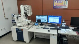
The institutional microscopy platform of materials characterization hosts the scanning electron microscope (SEM) of the Concordia node.
Concordia Microscopists
Concordia Equipments
Equipment that are part of RQMEM
Other equipment (not affiliated with RQMEM)
Polytechnique Montreal / University of Montreal
Polytechnique Montréal and University of Montréal nodes hosts the microscopy platforms of the University of Montréal (Courtois Institute) and Polytechnique Montréal (Center for Microscopic Characterization of Materials)
Center for Microscopic Characterization of Materials (CM)2 , installed in Polytechnique Montreal, is expert in crystallography and metallurgy. The (CM)2 currently has the microscopes of the Polytechnique Montreal and University of Montreal nodes. A new transmission electron microscope will be installed in 2027.
Courtois Institute, at the University of Montreal, will soon welcome a new state of the art microscopie affiliated to Quebec Network of Electron Microscopy for Materials. Stay tuned to know more.
Polytechnique Montreal and University of Montreal Microscopists
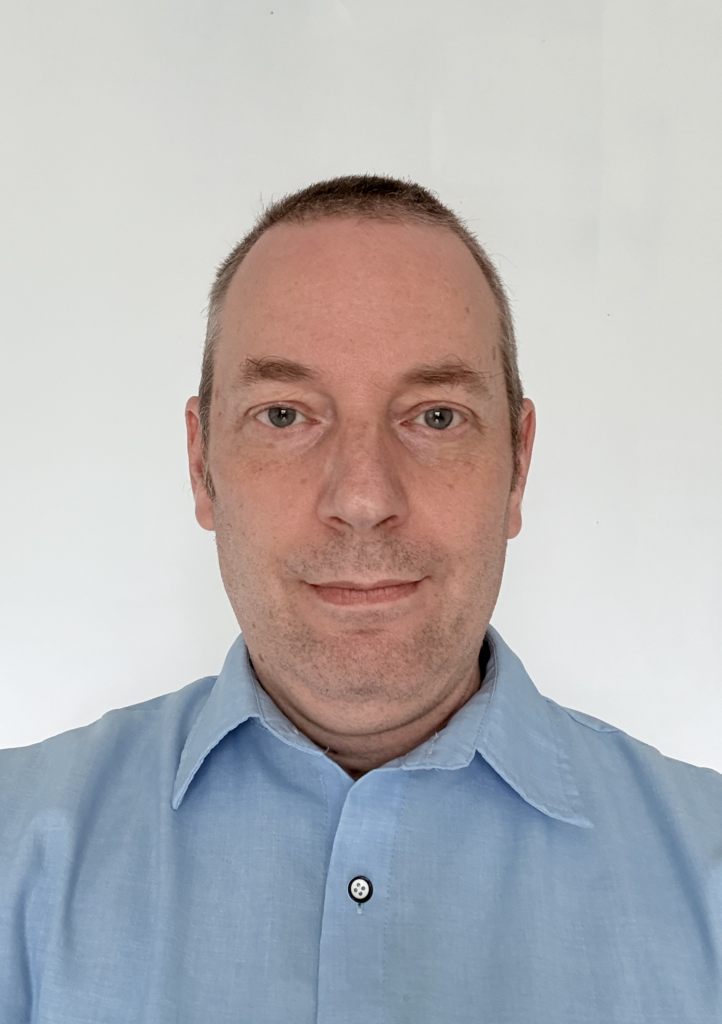
Titulaire d’une maîtrise en génie des matériaux, Philippe Plamondon est professionnel de recherche au Centre de caractérisation microscopique des matériaux, le (CM)2, à Polytechnique Montréal, où il travaille depuis 24 ans.
Au fil des années, il a acquis une vaste expérience pratique des différentes techniques de microscopie et de caractérisation disponibles au (CM)2. Ses activités se concentrent aujourd’hui principalement sur la microscopie électronique à balayage (MEB) et les techniques de faisceau ionique focalisé (FIB), notammentla reconstruction 3D par plasma FIB. En plus d’aider de nombreuses entreprises à relever leurs défis liés aux matériaux, il participe également à l’enseignement de la caractérisation microscopique à Polytechnique Montréal et contribue à la formation des étudiantes et étudiants aux tecniques de microscopie électronique.
Polytechnique Montreal and University of Montreal Equipments
Equipment that are part of RQMEM


University of Sherbrooke
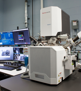
3iT.materials is home to a section dedicated to the RQMEM including the TEM, Ga FIB and a laboratory for sample preparation. This node is equipped for ultrahigh resolution with advanced spectroscopy like electron energy loss spectroscopy (EELS) and energy dispersion X-Ray spectroscopy (EDX).
University of Sherbrooke's Node microscopist, Frédéric Voisard, is expert in image analysis for segmentation, classification, metrology, industrial vision and artificial neural networks. He was trained in visualization and segmentation and in how to use Dragonfly (COMET).
University of Sherbrooke Microscopists
University of Sherbrooke Equipments
Equipment that are part of RQMEM
Other equipment (not affiliated with RQMEM)
Laval University
Laval University of an important pole in material science in Quebec. Many microscopy platforms are at University Laval, on and off campus.
Laval University Microscopists
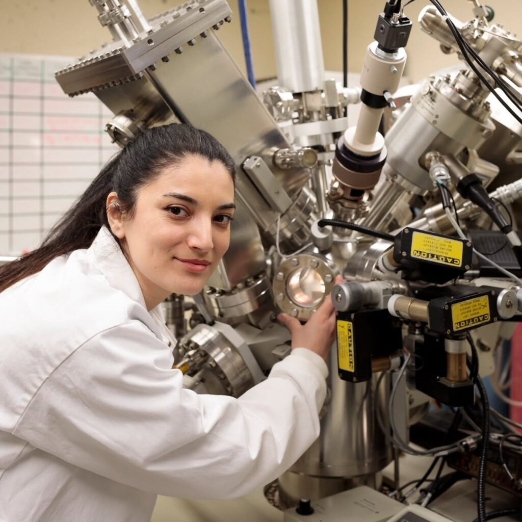
Samira Ravanbakhsh est professionnelle de recherche au Centre de recherche du CHU de Québec–Université Laval, au sein du BIMlab dirigé par le professeur Marc-André Fortin. Son expertise comprend la caractérisation des biomatériaux et des nanoparticules, l’analyse des surfaces et des interfaces ainsi que la microscopie électronique avancée. Depuis 2025, elle contribue de façon importante à la mise en place du service de cryo-microscopie électronique en transmission (cryo-TEM) au Centre de recherche du CHU de Québec–Université Laval (collaboration entre le BIMlab et la Plateforme de Bio-imagerie). Ses travaux portent notamment sur la visualisation et la caractérisation de matériaux biologiques et de matériaux mous, tels que les vésicules extracellulaires, les liposomes, les particules de type viral, les hydrogels, les nanoparticules et les tissus tumoraux, à l’aide de la cryo-microscopie électronique et de techniques complémentaires de caractérisation et de méthodes avancées d’analyse d’images.
Samira détient un doctorat en génie des matériaux de l’Université Laval, avec une spécialisation en biomatériaux. Au cours de ses études doctorales, elle a travaillé sur la modification de surfaces et la caractérisation d’endoprothèses cardiovasculaires et d’implants osseux. Elle a également reçu une formation spécialisée à la plateforme de recherche en microscopie électronique (FEMR) de l’Université McGill en microscopie électronique en transmission à haute résolution, appliquée à la caractérisation de nanoparticules à base de terres rares. En parallèle de ses activités de recherche, elle est chargée d’enseignement, elle est de plus chargée d’enseignement à l’Université Laval pour des cours dans le domaine des biomatériaux, notamment en nanobiomatériaux pour la théranostique et en modification de surface des biomatériaux.

Alexandre Bastien : Trained in physics at University of Sherbrooke (2008, B. Sc.) and at the Centre for optics, photonics and lasers of University Laval (2010, M. Sc.), he then worked for 8 years as a research professional in confocal microscopy and 2 years as a physics technician. Since 2021, he is responsible for the imaging and microscopy platform IBIS at University Laval. While hosted in a life science institute, this platform welcomes many groups and projects in material science. There is a TEM, a SEM, a sample preparation and ultra-microtomy service, a histology service and many optical microscopes with high resolution. Among the most interesting materials, Alexandre likes carbon nanotubes (TEM) and wood fibers extruded by electrofiling (SEM) best.
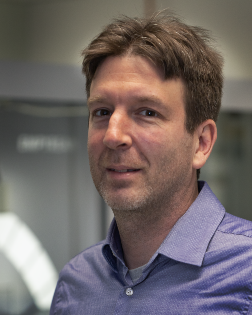
Diplômé de l’École Polytechnique en génie physique (2004) et titulaire d’un doctorat de l’INRS-EMT (2010), Nicholas Larouche a été professionnel de recherche à INRS-EMT jusqu’en 2015 (recherche et développement de catalyseurs non-nobles pour les piles à combustible à hydrogène) avant d’aller dans l’industrie (développement et mise à l’échelle de procédé de production de nanomatériaux de carbone chez Raymor). En 2023, il a rejoint la plateforme de caractérisation du Microlab à l’Université Laval à titre de responsable de travaux pratiques et de recherche afin de répondre aux besoins de la clientèle académique, gouvernementale et industrielle entourant les analyses par microscopie électronique à balayage. Fort de multiples expériences sur la synthèse de matériaux et le développement de procédés, il possède une expertise approfondie en caractérisation et une vision large qui aident à répondre aux besoins de la clientèle avec professionnalisme.
École de Technologie Supérieure
The ETS is home to the Network's Ion Miller for sample preparation. It is installed in a platform with other equipments for the caracterization and fabrication of materials.
ETS microscopists
ETS equipments
Equipment that are part of RQMEM
Other equipment (not affiliated with RQMEM)