
Published on December 2, 2025
On October 16, 2025, the University of Sherbrooke Node held the inauguration of its electron microscopy platform, 3IT.Materials. Several RQMEM researchers were present at the Interdisciplinary Institute for Technological Innovation (3iT) for the official lauch of the focused ion beam (FIB) scanning electron microscope and the transmission electron microscope (TEM).
These microscopes, made available because of the collaboration between different nodes of the network, the partner universities, the Canadian Foundation for Innovation (CFI) and the Ministère de l'Économie, de l'innovation et de l'énergie, are accessible to all members of the RQMEM to help research in advanced materials, train highly qualified students and professionals, et stimulate collaboration within the Network and the province of Quebec.
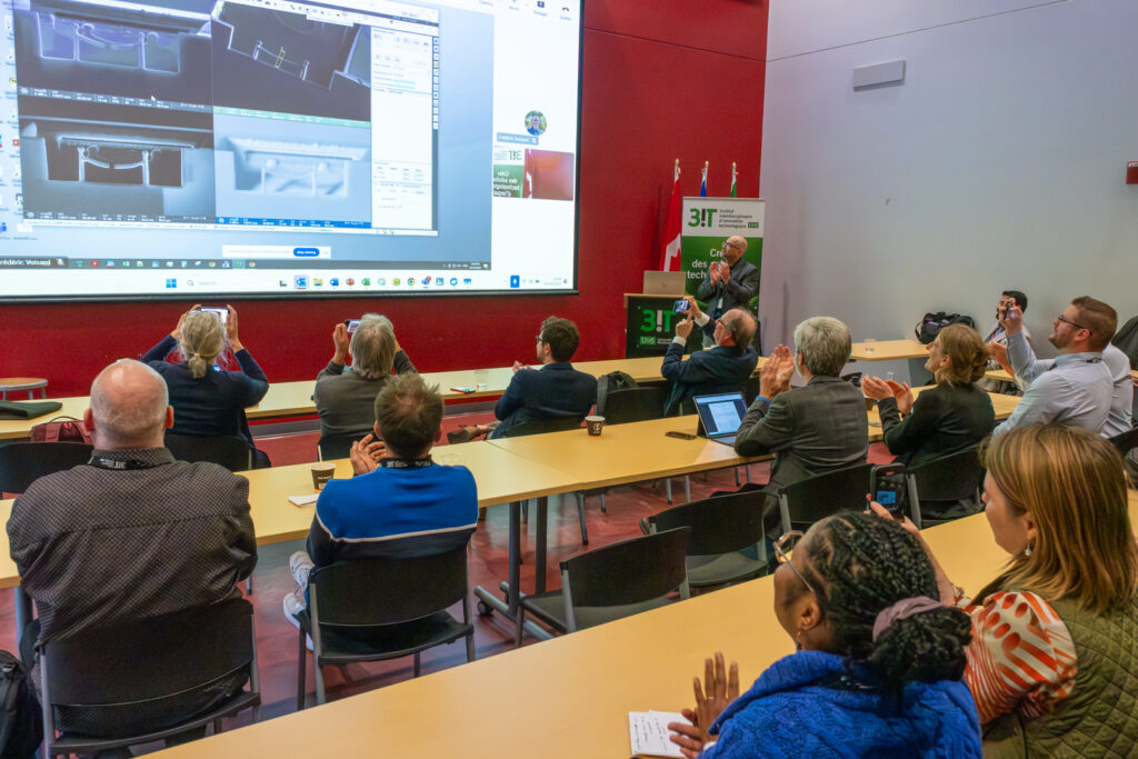
The present members, including node directors and node microscopists, assisted to a symbolistic chord cutting, directly from the transmission electron microscope.
The 3IT.Material platform benefited from a funding of millions of dollars to build 1 000 m2 of net laboratory surface, 300 m2 of office surface for the highly qualified personal and a new landing stage. Other than the room dedicated to the transmission electron microscope, a room for the FIB and a sample preparation room (cutting, polishing, electropolishing, light microscopy), some room is still available for future equipment for material characterisation.
The 3IT.Materials is now a pillar of microelectronics, nanotechnology, quantum and advanced materials, energy devices research, to name a few.
The microscopes inaugurated make many analysis possible in high resolution imagery, crystallographic analysis and elementar and chemical phase analysis.
The equipments within the microscopy platform of 3IT.Materials
Scanning electron microscope and focalised ion beam (SEM/FIB) allows the surface morphology observation, ion beam cuts, tomography but plan cuts as well as chemical analysis by EDS. This Scios 2 SEM-FIB from ThermoFisher is powered by a Schottky FEG canon. The detectors and spectrometers allows the analysis of secondary electrons, filtered secondary electrons, and backscattered electrons as well as X ray spectroscopy for a composition or cartography of the materials. This equipment allows micromanipulation like cutting at the microstructure scale or observing under a covered or hidden surface. It also allows to prepare thin electron transparent films for the transmission electron microscope.
The transmission electron microscope (TEM) can be used to visualize materials to the atomic scale, for electron diffraction and chemical mapping (EDS or EELS). Spectra 200 from ThermoFIsher has a spherical abheration corrected cold FEG canon. It allows a resolution as low as 60 pm. The sample holder allows in situ analysis up to 1 atm or 1 000 oC with or without an electronic bias imposed by microelectrodes. This sample holder allows observation during the micro- and nanostructure evolution in reactive conditions.
Images can be observed using a 4K Falcon camera (in imagery or diffraction mode), four EDX spectrometers surrounding the sample, an electron energy loss spectrocopy camera (K2-GIF-Continuum EELS) and segmented transmission detectors allowing differential contrast imagery (on bright field, dark field, annular or wide angle annular).
For more information on the equipments available within the RQMEM, consult our instrumentation page. More informations on how to access equipments is available on our page on RQMEM"s microscope access of RQMEM.