




A node is an institution of higher education that supports the Quebec Network of Electron Microscopy for Materials and where there is one or more microscopy platform. Some nodes are part of the institutions who originally submitted for funding with the Canadian Foundation for Innovation, in 2017, which are University of Sherbrooke, McGill University, Polytechnique Montréal, University of Montréal, Concordia University, Laval University and École de technologie supérieure. Other partner institutions can be added as RQMEM nodes with the approval of the RQMEM board of directors.
McGill
FEMR (Facility for Electron Microscopy Research) and MEMRG (McGill Electron Microscopy Research Group) are microscopy platforms members of the Quebec Network of electron microscopy for materials and bring together a diversified expertise with many microscopists collaborating with the Network.
FEMR is a platform that serves material science and biological science with expertise in tomography, molecular reconstruction and 3D vizualisation.
MEMRG holds the expertise for X ray quantification, materials and batteries analysis.
McGill Microscopists
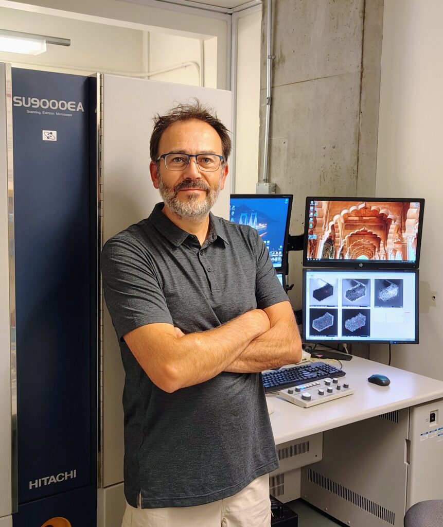
Nicolas Brodusch is a research assistant at McGill and affiliated with theMcGill Electron microscopy Research Group, MEMRG, www.memrg.com), directed by professor Raynald Gauvin. As well as managing the equipment's operations and coordinating the student's research projects in the group, he develops innovating techniques, in the sample preparation field as well as in diverse material characterization by scanning electron microscopy.
Nicolas obtained a Technology University Diploma (DUT), with Physical Measures option, à Université Paris XI at Orsay, France, in 1997. After perfectionning analysis and research techniques as a laboratory technician in different laboratories, he obtained his masters in Materials Science and Engineering by validating professional experience at the University Paris-Saclay, Orsay, France, in 2018. During the sixteen years that he spent at MEMRG, Nicolas pushed the limits of scanning electron microscopy (SEM) to improve the imaging resolution and image quality, but also to use the detectors equiped on the instruments to their full potential. His work was internationally recognized by numerous scientific publications and the attributing an award at the Microscopy & Microanalysis conference in Hartford, CT, in 2014 for the article "Dark-field imaging of thin specimens with a forescatter electron detector at low accelerating voltage", and many invitationsa s a guest presenter in conferences in the last few years.
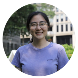
Wanting He is currently a postdoctoral fellow at McGill University and is affiliated with the Facility for Electron Microscopy Research (FEMR). RQMEM is proud to have access to her expertise in transmission electron microscopy (TEM), electron energy-loss spectroscopy (EELS), electron tomography, and nanomaterials characterization (i.e. plasmonics materials).
She received her B.S. in Materials Science and Engineering from Beihang University and her M.S. in Materials Engineering from the University of Southern California. She earned her Ph.D. in Materials Science from the Institut National de la Recherche Scientifique (INRS), where her research focused on time-resolved ultrafast transmission electron microscopy (UTEM).

Angel Valdez is a Research Associate at the Facility for Electron Microscopy Research (FEMR) at McGill University, where heI develops and applies electron microscopy techniques for materials characterization and structural analysis. His expertise includes Microcrystal Electron Diffraction (MicroED), scanning transmission electron microscopy (STEM), energy-dispersive X-ray spectroscopy (EDS), electron energy-loss spectroscopy (EELS), electron tomography, and cryogenic electron microscopy. He is particularly interested in developing new workflows that expand the capabilities of electron microscopy and make advanced techniques more accessible to the scientific community.
Angel Valdez holds a Ph.D. in Energy and Materials Science from INRS in Quebec. He subsequently joined McGill University, where he currently works as a Research Associate at FEMR. His research focuses on developing innovative electron microscopy methodologies. In addition to research, he enjoys supporting users providing technical and training support, teaching microscopy techniques, and organizing workshops and presentations that help researchers make the most of advanced microscopy instrumentation.
McGill equipments
Equipment that are part of RQMEM


Other equipment (not affiliated with RQMEM)
Concordia
The institutional microscopy platform of materials characterization hosts the scanning electron microscope (SEM) of the Concordia node.
Concordia Microscopists
Concordia Equipments
Equipment that are part of RQMEM
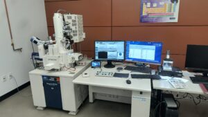
Other equipment (not affiliated with RQMEM)
Polytechnique Montreal / University of Montreal
Polytechnique Montréal and University of Montréal nodes hosts the microscopy platforms of the University of Montréal (Courtois Institute) and Polytechnique Montréal (Center for Microscopic Characterization of Materials)
Center for Microscopic Characterization of Materials (CM)2 , installed in Polytechnique Montreal, is expert in crystallography and metallurgy. The (CM)2 currently has the microscopes of the Polytechnique Montreal and University of Montreal nodes. A new transmission electron microscope will be installed in 2027.
Courtois Institute, at the University of Montreal, will soon welcome a new state of the art microscopie affiliated to Quebec Network of Electron Microscopy for Materials. Stay tuned to know more.
Polytechnique Montreal and University of Montreal Microscopists
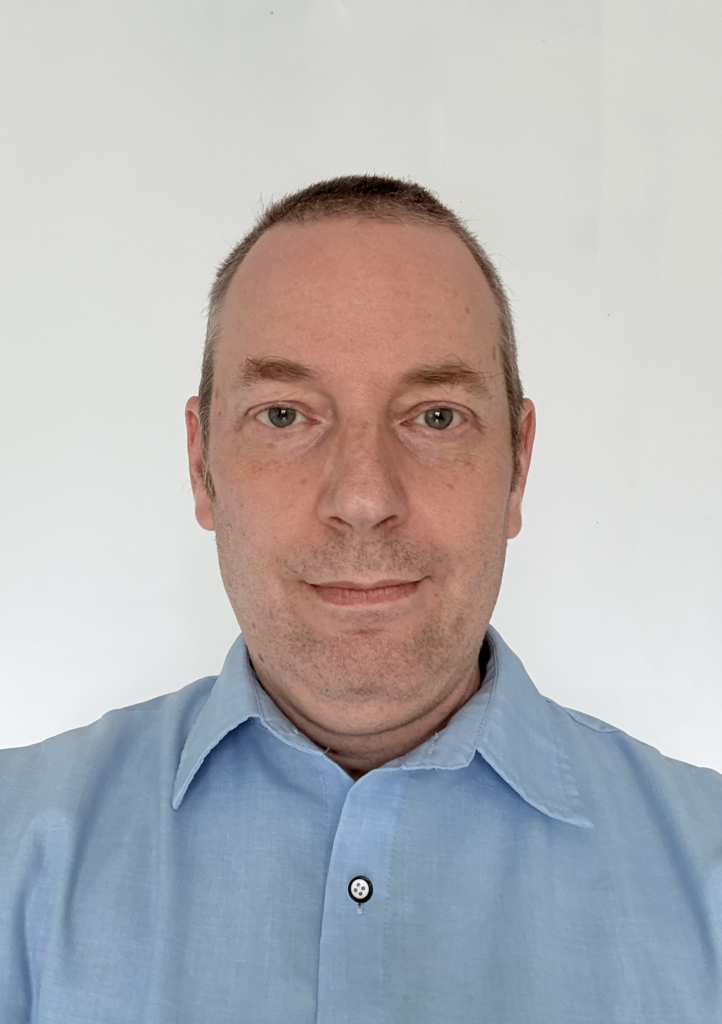
Master in materials engineering, Philippe Plamondon is a research associate at Centre de caractérisation microscopique des matériaux, the (CM)2, at Polytechnique Montreal, where he has been working for 24 years.
Accross the years, he acquired great practical experience on different microscopy and characterization techniques available at (CM)2. His activities are now mainly on scanning electron microscopy (SEM) and focalised ion beam techniques (FIB), especially the 3D reconstruction by plasma FIB. AS well as helping numerous companies to solve their materials challenges, he participates to the training on microscopic characterization at Polytechnique Montreal and the training of students on electron microscopy techniques.
Polytechnique Montreal and University of Montreal Equipments
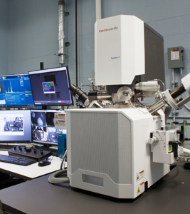
Equipment that are part of RQMEM


University of Sherbrooke
3iT.materials is home to a section dedicated to the RQMEM including the TEM, Ga FIB and a laboratory for sample preparation. This node is equipped for ultrahigh resolution with advanced spectroscopy like electron energy loss spectroscopy (EELS) and energy dispersion X-Ray spectroscopy (EDX).
University of Sherbrooke's Node microscopist, Frédéric Voisard, is expert in image analysis for segmentation, classification, metrology, industrial vision and artificial neural networks. He was trained in visualization and segmentation and in how to use Dragonfly (COMET).
University of Sherbrooke Microscopists
University of Sherbrooke Equipments
Equipment that are part of RQMEM
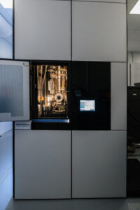
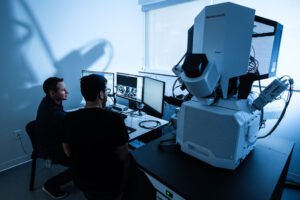
Other equipment (not affiliated with RQMEM)
Laval University
Laval University of an important pole in material science in Quebec. Many microscopy platforms are at University Laval, on and off campus.
Laval University Microscopists
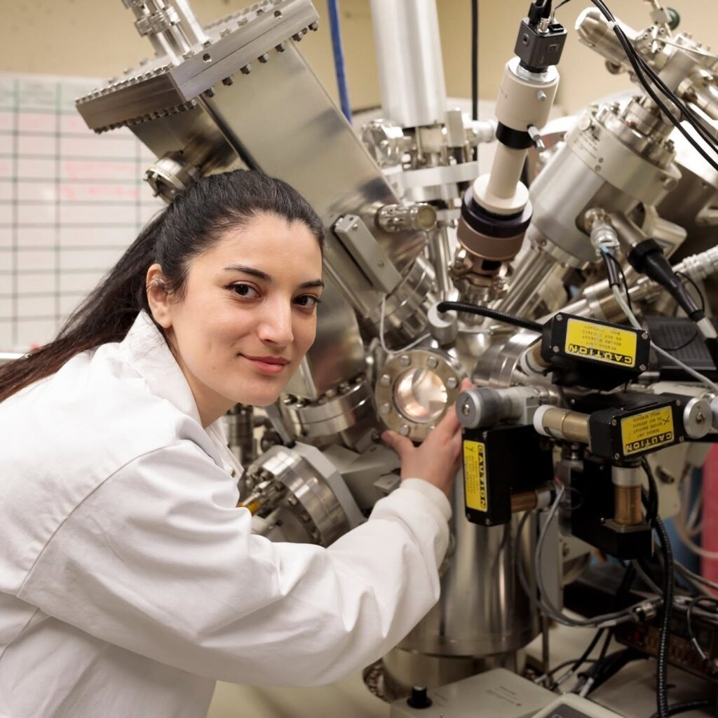
Samira Ravanbakhsh is a research associate at the CHU-Québec-University research center, in BIMlav directed by Marc-André Fortin. Her expertise includes biomaterial and nanoparticles characterization, surfaces and interfaces analysis and advanced electron microscopy. Since 2025, she contributes to the implementation of cryo-transmission electron microscopy (cryo-TEM) service at the CHU de Quebec - Université Laval research center (collaboration between BIMlab and bio-imaging platform). Her work is focused on the vizualisation and characterization of biological and soft materials like extracellular vesicules, liposomes, viral particles, hydrogels, nanoparticles and tumor materials, using cryo-electron microscopy and complementary characterization and advanced image analysis techniques.
Samira has a PhD in materials engineering from Université Laval, with a specialisation in biomaterials. During her doctoral years, she worked on modifying surfaces and characterization of cardiovascular endoprothesis and bone grafts. She also trained at the electron microscopy platform, FEMR, at McGill University in transmission electron microscopy with high resolution, applied to the characterization of nanoparticles made of rare earth materials. In addition to her research activities, she is a teaching assistant at Université Laval for courses in biomaterials, including in nanobiomaterials for theranostic and surface modification of biomaterials.

Alexandre Bastien : Trained in physics at University of Sherbrooke (2008, B. Sc.) and at the Centre for optics, photonics and lasers of University Laval (2010, M. Sc.), he then worked for 8 years as a research professional in confocal microscopy and 2 years as a physics technician. Since 2021, he is responsible for the imaging and microscopy platform IBIS at University Laval. While hosted in a life science institute, this platform welcomes many groups and projects in material science. There is a TEM, a SEM, a sample preparation and ultra-microtomy service, a histology service and many optical microscopes with high resolution. Among the most interesting materials, Alexandre likes carbon nanotubes (TEM) and wood fibers extruded by electrofiling (SEM) best.
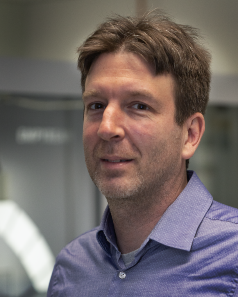
With a diploma from Polytechnique Montreal in physics engineering (2004) and a PhD from INRS-EMT (2010), Nicholas Larouche was a research associate at INRS-EMT until 2015 (research and development of non-noble catalysts for hydrogen combustion batteries) before going into the industry (development and scale up of production process for carbon nanomaterials at Raymor). In 2023, he joined the characterization platform Microlab at Université Laval as practical work and research coordinator to help with the academic, governement, and industrial clients regarding the scanning electron microscopy (SEM). Strong with multiple experience in materials synthesis and process development, he has a strong expertise in characterization and a large vision that helps meet the needs of the clients with professionalism.
École de Technologie Supérieure
The ETS is home to the Network's Ion Miller for sample preparation. It is installed in a platform with other equipments for the caracterization and fabrication of materials.
ETS microscopists
ETS equipments
Equipment that are part of RQMEM
Other equipment (not affiliated with RQMEM)