

Crossover Between Industry and Research in Aquaculture
Imaging demos
First, the group learned about generral imagery by observingthe morphology of a scallop by scanning electron micrscopy. The instrument used was the SEM Hitachi SU3500 with variable pressure mode (SP-SEM) to improve the sample's condution without previously addind metal.
In a second room, we pushed the analysis of microsctructre and composition of a scallop on the SEM Hitachi SU8000. Two samples were observed, one polished which allowed to observe the internal structure and the fine details of calcium carbonate grains, and a fractured sample allowing to access the microstructure and the 3D internal organization of grains.
During this analysis, the secondary electron detector (SE) was used to observe the grain morphology, especially the lamellar shape, and the backscattered electron detector (BSE) allowed to visualize the diferent chemical compositions and the grain orientation on the polished sample due to channeling effect. THe group could discover a zone with a different morphology at the center of the shell showing a distinctive grain distribution.
A complete complementary EDS sanalysis confirmed the sample was mostly made of calcium (Ca), carbon (C), and oxygen (O), corresponding to the expected calcium carbonate composition.


From left to write, we observe teh EBSD image of the scallop three times, then the fractured scallop, and the polished scallop, during a microstructure analysis.
Lastly, the group moved to a third room to do the crystallographic analysis of a previously polished scallop with perfectly plane surface. These conditions are essential for a robust EBSD analysis. The instrument used was the SEM Hitachi SU8230 equipped for EBSD (Electron Backscatter Diffraction). This analysis identified the different phases in the calcium carbonate (confirmed during the previous analysis).
Two phases were identified : calcite and aragonite. The calcite was the main phase, made of longiform lamellar bunches with various orientations in the shell. Aragonite was a finer zone of 100 µm thickness going through the sample. It has a distinctive cristalline structure and a different grain composition.
The presentations were as varied as the attendees. From the natural History of bivalve mollusks (Frederic Marin, Université de Bourgogne) to theFabrication of dynamic bio-interfaces in the mussel byssus (Matthew Harrington, McGill), were also discussed the impact of anthropogenic sound on mussel shell integrity (Réjean Tremblay, UQAR), and the impact of marine heatwaves on Mytilus edulis (Guillaume Durier, UQAR), as well as the potential impact of metal pollution on shell strenght in Pecten Maximus (Roland Kröger, University of York).
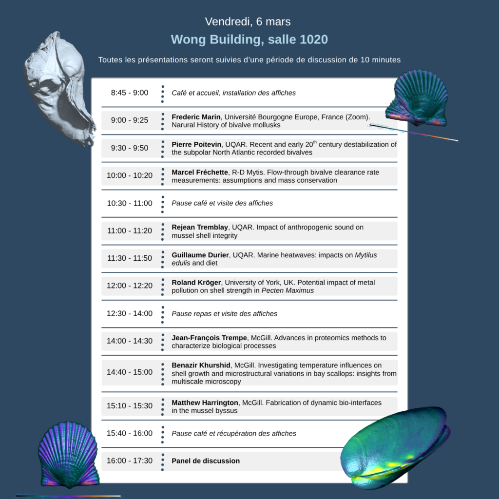
Other presentation discussed the recent and early 20th century destabilization of the subpolar North Atlantic recorded bivalves (Pierre Poitevin, UQAR), assumptions and mass conversation about the Flow-through bivalve clearance rate measurements (Marcel Fréchette, R-D Mytis), advances in proteomics methods to characterize biological processes (Jean-François Trempe, McGill), and a study of the temperature influences on shell growth and microstructural variations in bay scallops (Benazir Khurshid, McGill).

Apart from oral presentation, a poster session allowed some presenters to discuss their research in a more casual setting, favoring exchange with the pairs.
On the left, we can see these discussion around one of the posters.
The poster subjects were varied, talking about custom detox systems for artificial seawater, characterizing the role of novel zinc-binding proteins in mussel adhesive curing, proteinaceous thread assembly of zebra and quagga (dreissenidae), illuminating the neural network of the Mytilus edulisthread complex, and the X-ray characterization of Fe and V coordination complexes for mussel byssus reinforcement.
A symposium bringing together scientists from many fields is the perfect opportunity to diversify ideas and solve problems with innovating, maybe out of the box, solutions. This symposium was funded by Réseau Québec Maritime andMcGill Sustainability Systems Initiative.
The RQMEM is proud to participate to the organizaiton of this event and hopes that the exchanges between pairs were fruitful. Networking is part of the RQMEM's mission, to obtain our help with planning similar events, do not hesitate to communicate with us by sending an email to rqmem@usherbrooke.ca.